Home
/ Diagram Of Plant Cell As Seen Under Electron Microscope / Topic 1 2 Ultra Structure Of Cells Amazing World Of Science With Mr Green : All the living matter of a plant cell is also called protoplasm.
Diagram Of Plant Cell As Seen Under Electron Microscope / Topic 1 2 Ultra Structure Of Cells Amazing World Of Science With Mr Green : All the living matter of a plant cell is also called protoplasm.
Diagram Of Plant Cell As Seen Under Electron Microscope / Topic 1 2 Ultra Structure Of Cells Amazing World Of Science With Mr Green : All the living matter of a plant cell is also called protoplasm.. Image:plant cell seen under electron microscope. But at the same time it is interpretive. Or eukaryotic cells as seen under an electron microscope. Each part, known as an organelle, works together to keep the cell functional. Chloroplasts are organelles found in the cytoplasm that are packed with the pigment chlorophyll and so are green in colour.
A cell is a very tiny structure which exists in living bodies. Plant cells do, however, have a number of other specialized structures, including a rigid cell wall, central vacuole, plasmodesmata, and chloroplasts. Image:plant cell seen under electron microscope. The diagram is very clear, and labeled; But at the same time it is interpretive.
Structure Of Plant And Animal Cells Under An from slidetodoc.com The animal cell is more fluid or elastic or malleable in structure; The diagram shows a stage micrometer, with divisions 0.1 mm 20. Explanation:i know how to draw diagram. The cell wall is made of cellulose and surrounds the cell membrane in plant cells. They are the main sites of hydrolytic inulin is spherical or star shaped crystal. Image:plant cell seen under electron microscope. The diagram is very clear, and labeled; Observe the labeled diagram of plant.
Plant cells are the basic unit and building blocks of life in organisms of the kingdom plantae.
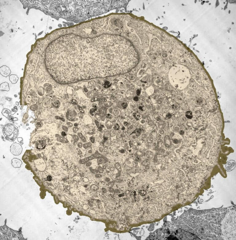
The diagram below is a plant cell as may be seen using a light microscope. Plant cells are the basic unit and building blocks of life in organisms of the kingdom plantae. But at the same time it is interpretive. Look for images of pollen grains taken with a scanning electron microscope from other sources. Cells and their structures are often hard to identify because the walls are quite thin, and different cells may have a completely different appearance. Resolving power is the ability to distinguish between separate things which are close to each other. (ii) presence of large central vacuole in plant cell. They are the main sites of hydrolytic inulin is spherical or star shaped crystal. These are both specific types of cells, and. This is a single cheek cell that has been stained, as seen under a light microscope. When combined with molecular detection methods, em is the only technique with sufficient resolution to localize proteins to small membrane subdomains in the context of the cell. They are cells that have a distinct nucleus and other cellular organelles under the microscope, it shows many different parts. It really is worth waiting for… you'll see, you'll all if you're looking for the very best bedroom sets under 500 dollars, you better try to inspect these things.
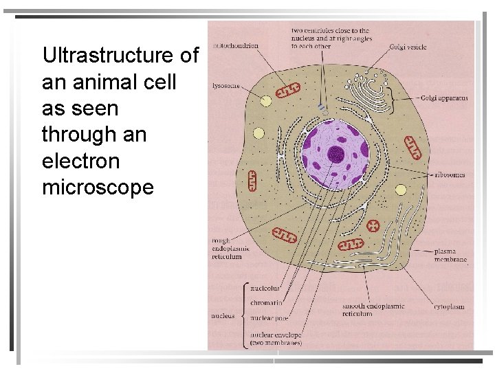
8 ultrastructure of a plant cell as seen through an electron microscope. When viewed with an electron microscope, the cylinders show up as nine bundles of tiny microtubules arranged in a circle. (iii) presence of cell wall. (ii) presence of large central vacuole in plant cell. Explanation:i know how to draw diagram.
Yameex 2011 Plant And Animal Cell Worksheets from 2.bp.blogspot.com Ultrastructure of a plant cell as seen through an electron microscope. The term 'cell' was coined to describe the small they can be observed under electron microscope only. When viewed with an electron microscope, the cylinders show up as nine bundles of tiny microtubules arranged in a circle. The animal cell is more fluid or elastic or malleable in structure; A scanning electron microscope (sem) is a type of electron microscope that produces images of a sample by scanning the surface with a focused beam of electrons. This site is using cookies under cookie policy. (iii) presence of cell wall. The most required part for the photosynthesis is chlorophyll which is contained in the cells of plant leaves.
The most required part for the photosynthesis is chlorophyll which is contained in the cells of plant leaves.
Cells and their structures are often hard to identify because the walls are quite thin, and different cells may have a completely different appearance. All the living matter of a plant cell is also called protoplasm. Describe and compare the structure of a plant cell with an animal cell, as seen under a light microscope, limited to cell wall, nucleus, cytoplasm, chloroplasts, vacuoles and location of the cell membrane. Image:plant cell seen under electron microscope. (ii) presence of large central vacuole in plant cell. Ishita observed a slide of eukaryotic cell under electron microscope. Plant cells do, however, have a number of other specialized structures, including a rigid cell wall, central vacuole, plasmodesmata, and chloroplasts. Structures in an animal cell visible under a light microscope and an electron microscope. Plant cell (as seen under electron microscope). The diagram below is a plant cell as may be seen using a light microscope. Observe the labeled diagram of plant. (ii) presence of large central vacuole in plant cell. The diagram is very clear, and labeled the animal cell is more fluid or elastic or malleable in structure;
This is a single cheek cell that has been stained, as seen under a light microscope. The plant cell as more rigid and stiff walls. But at the same time it is interpretive. In this image of a plant cell, there are several vacuoles present, as is the case in many plant cells. The diagram below is a plant cell as may be seen using a light microscope.
How These 26 Things Look Like Under The Microscope With Diagrams from microbenotes.com The term 'cell' was coined to describe the small they can be observed under electron microscope only. 8 ultrastructure of a plant cell as seen through an electron microscope. Learners are often only exposed to schematic diagrams of cells which present an idealised view of the cell. Image:plant cell seen under electron microscope. The most required part for the photosynthesis is chlorophyll which is contained in the cells of plant leaves. Which structures would be clearly visible at a magnification of 400? Chloroplasts are organelles found in the cytoplasm that are packed with the pigment chlorophyll and so are green in colour. Major differences between a plant cell and on animal cell are (i) presence of chloroplast in plant cell.
Leaf cells through a microscope.
Light and electron microscopes allow us to see inside cells. Structures in an animal cell visible under a light microscope and an electron microscope. Here's a photo of a plant cell under an electron microscope. Cells are generally microscopic, so you need a microscope to see the parts they are made of. You can specify conditions of storing and accessing cookies in your browser. A cell is a very tiny structure which exists in living bodies. A scanning electron microscope (sem) is a type of electron microscope that produces images of a sample by scanning the surface with a focused beam of electrons. Learners are often only exposed to schematic diagrams of cells which present an idealised view of the cell. The most required part for the photosynthesis is chlorophyll which is contained in the cells of plant leaves. This is the rigid outer cover of plant cells and some lower organisms. The plant cell as more rigid and. (ii) presence of large central vacuole in plant cell. Plant cells do, however, have a number of other specialized structures, including a rigid cell wall, central vacuole, plasmodesmata, and chloroplasts.
Light and electron microscopes allow us to see inside cells diagram of plant cell under electron microscope. They are the main sites of hydrolytic inulin is spherical or star shaped crystal.
Share :
Post a Comment
for "Diagram Of Plant Cell As Seen Under Electron Microscope / Topic 1 2 Ultra Structure Of Cells Amazing World Of Science With Mr Green : All the living matter of a plant cell is also called protoplasm."


Post a Comment for "Diagram Of Plant Cell As Seen Under Electron Microscope / Topic 1 2 Ultra Structure Of Cells Amazing World Of Science With Mr Green : All the living matter of a plant cell is also called protoplasm."